


Onychomycosis is a kind of mushroom lesion that only affects the nail surface.In order to prevent the spread of infections in good time, you should know what the nail fungus looks like, since the therapeutic effect is achieved faster when treatment begins at an early stage of the infection.Most of the time, the disease occurs in older people due to weak immunity, but the risk of maintaining an illness concerns everyone.There is not a single classification of mushroom nail damage;In medical practice, it is common to distinguish them at the place of localization and depth into which they penetrate.The infection is also grouped depending on the type of pathogen.
Types of pathogens and primary signs
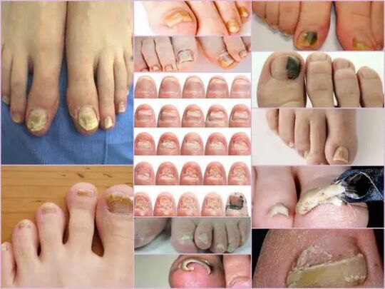
The symptoms of nail fungus in the initial stage are easy to determine directly by the condition of the nail.This sign is most informative, since onychomycosis always manifests itself in the form of changes in the color of the surface, the deformation of the bed, peeling and all external changes.The latter are expressed by roughness, the formation of grooves and cracks as well as through a well -being and general violation of the nail.
The main sign of a healthy nail is pink and transparency.Onychomycosis at every stage is characterized by the turbidity of the nail and the change in color too yellow, brownish or greenish-black.In an advanced level, the surface can achieve a black shade against the background of almost complete destruction.
External signs of infection with a mushroom depend on the type of infected pathogen.In medical practice, the following possible lesions are differentiated:
- Candida infection with a mushroom that is expressed directly at the base of the box by unloading the nail.Candidiasis of the roles around the nail will be characteristic of candidiasis.This version of the complication can either have a bacterial source in the form of streptococci or staphylococci or be expressed in the form of medium or psoriasis plaques;
- Dermatophytes of the Trichophyton -rubrum type.In this case, the infection is penetrated directly from the free edge of the nail.The first symptoms of such a pathogen are the appearance of a yellowish stain on the surface.The nail collapses in the area of neoplasm and the stain itself tends.The common place of the localization of the neoplasm lies along the plate, parallel to the nail rollers, in this case the infection is described as distal acateral.There is another form of defeat by this pathogen - distal, in which the part of the part of the hole appears, mainly in the middle of the free edge.
Important!
This type of mushroom most often occurs on the thumb of the legs and gradually turns the nails into a loose yellowish mass.In the absence of proper treatment, the disease flows into hyperkeratosis.Due to the spread of the infection, the nail plate is completely destroyed by the scope.
- Dermatophytes such as trichophyton mentagrophytes.Onychomycosis with such suggestions is most often based on the nails of the thumb, less often than the little fingers.An infection with a fungus of this kind requires therapy not only of the nail, but also the feet due to the rapid spread of the pathogen.A symptomatic illness can be like Leonkonichia, a disease common in medical practice.The main signs are white spots that occur on different parts of the nail.Neoplasms are differentiated by a non -standard shape and different sizes.It is easy to distinguish the mushroom from Leckonichie - in the latter case, the spots are an aerial bar cumulation that is not observed with mushroom damage.
- Mold.This damage option is much less common than a candidal or dermatophytic form.The main sign of such an infection - the surface of the plate gets a dark, almost black shade.Not the entire finger can be fully infected, but only part of the nail plate.The first signs of nail fungus on the legs of this type are a sharp change in color.Onychomycosis can develop in the form of a longitudinal strip made of black or dark green color against the background of the rest of the pink part of the nail.
Diagnostics after change
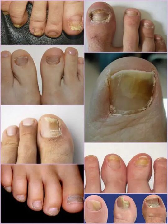
It is not difficult to notice a nail fungus on the legs even in the primary stages of infection, since an infection of this kind of the first day of the lesion manifests itself quite actively.Instead of the usual transparent nails of light pink in the patient, a significant surface deformation and a general change in the condition are observed.The affected area has a mat, matt yellowish color, which mainly occurs on the thumb.The type of mushroom and the degree of damage determine the factors.
In the first stage, the mushroom damage on the legs on the legs is the appearance of small focal points.The thickening of the plate and the keratization of the bed under the nail is characteristic.This level is accompanied by such a phenomenon such as partial detachment and the unloading of the nail plate, which serves as a source of infection for healthy people.
Despite the active thickening of the plate, the constant grinding can be observed regardless of the current factors.The characteristics of each stage and the symptoms of the mushroom on the legs depend directly on the type of pathogen.
Depending on the changes with the nail plate, three options for onychomycosis are differentiated:
- Hypertrophic;
- Normotrophic;
- Atrophic.
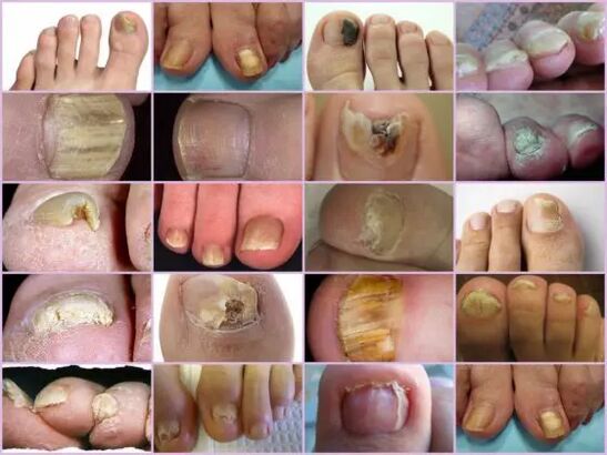
In the first case, there is a sharp change in the shadow of the nail plate, its destruction along the edges and the deformation of the surface of the plate.The nail thickens so much that it causes symptoms and painful sensations when walking.The normotrophic mycosis of nails on the legs differs by the presence of a healthy shine, but the plate itself gets something yellow on which stains arise.With an atrophic type of damage, brown and gray focal points form on the surface of the nails.Thanks to them, it is possible to precisely determine the location of the pathogen.
Important!
The atrophic or onycholithic nail fungus is characterized by thinning the nail plate and not by its growth as in other cases.The areas in which the pathogen is localized tends to tend.The lack of proper treatment leads to an advanced level - the complete rejection of the nail plate.
Localization classification
Another sign with which you can separate the mushroom damage on the nails of the toes depends directly on the position of the focus on the nail plate.This also includes the depth of the pathogen, with which you can in turn determine the approximate duration of the upcoming therapy and the chances of a quick recovery.
Mushroom diseases of the legs at the location of the localization are divided into the following groups:
- Onychomycosis is a white surface type - the appearance of many whitish spots on the surface of the nail plate.A fungal infection leads to the detachment of the skin in the places of the place of appearance of spots, where there is an active discharge of scales.The advanced stage leads to the full destruction and rejection of the plate;
- Distal - develops on the free border of the nail.The color change is initially observed in a small area of the plate, according to which there is an active expansion of its limits.The lesion is characterized by a yellowish-gray or brown color as well as a rough uneven surface and gradual peeling.
- Lateral onychomycosis has the stages of development similar to distal, but is only localized on the nail plate on the side.
- Total infection - complete infection of the entire surface;
- Proximal onychomycosis.This disease begins its activity from the cuticle, which is inflamed, and then the fungus quickly affects the nail, and the process itself begins with the appearance of a small white spot near the inflamed area of the periolous role.
The most common forms of mycosis of nails on the legs are lateral and distal, which is normally caused by dermatophytes.Such forms of damage such as proximal and white can act as a secondary disease that, for example, vich the disease of the immune system.Total damage to the nail with a mushroom should be seen as an advanced development stage of a mushroom under the nail.
Features of the mushroom under the microscope
Despite the presence of an impressive number of signs of onychomycosis, other diseases associated with skin problems and not contagious in nature for mushroom damage on the leg nails are accepted.You can only determine the exact diagnosis and the type of pathogen in the laboratory under a microscope, for which a scratch of organic material from the affected areas is carried out in a hospital.
The resulting biomaterials are proposed in an alkaline environment, according to which a multiple increase is carried out.If you examine the nail fungus under a microscope, you can see an active pathogen whose external shape determines its type, its distribution scale and an approximate degree of damage.On a nutrient medium, you can predict the approximate growth of the colonies, which not only makes it possible to make a precise diagnosis, but also to determine the restriction of the infection.
Important!
Since it is possible to only determine the presence of onychomycosis in one laboratory, you should not postpone a visit to the doctor with the slightest suspicion.The nail fungus develops quickly and the loss of time increases therapy.
What is an alarming signal?
The mushroom of nails on the legs is shown by certain symptoms, some of which are similar to some skin diseases.The following signs rather indicate an onychomyotic lesion that requires medical interventions:
- The appearance of yellow spots on the plate, its deformation, a change in the structure that was not observed earlier;
- The darkening of the plate, the loss of transparency, some photos of the nail fungus on the legs show a shadow near black, which is characteristic of forms of pathogens.
- Thickening and keratiization of the nail plate or vice versa thinning too sharp;
- Rejection of the nail out of bed, replacement of scales and crumbs;
- Swelling of the roller, hang over the plate, release liquid;
- The nails affected by the mushroom look inflamed regardless of the type of pathogen.
All of these symptoms indicate a high risk of infection.Some external symptoms of changing the structure of the nail plate can be the result of other diseases.The amount of calcium and iron in the body should be increased with increased crematriation, increased tuberosity means every infection of the body with purely white stripes and points, a lack of copper or zinc possible.Despite the fact that the nail fungus in the photo looks like a focal damage to the plate, whereby the color approaches closer to the yellow, this does not always indicate the infection.The yellowity can indicate various problems with stomach and liver.
It is not difficult to recognize onychomycosis by the fingers through external signs, despite the extensive classification of the disease.However, you can determine the exact type of the pathogen and the damage phase can only be in the laboratory.